
Anatomical model of a pregnant woman, polychrome boxwood, France or Germany, circa 1670
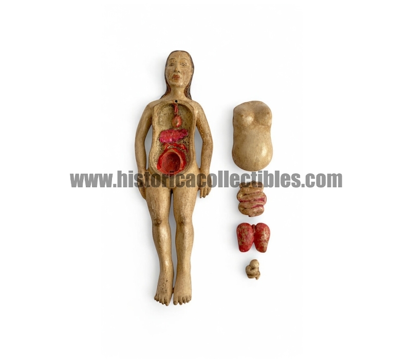
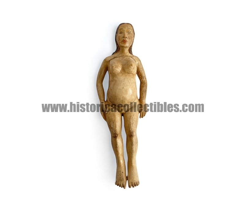
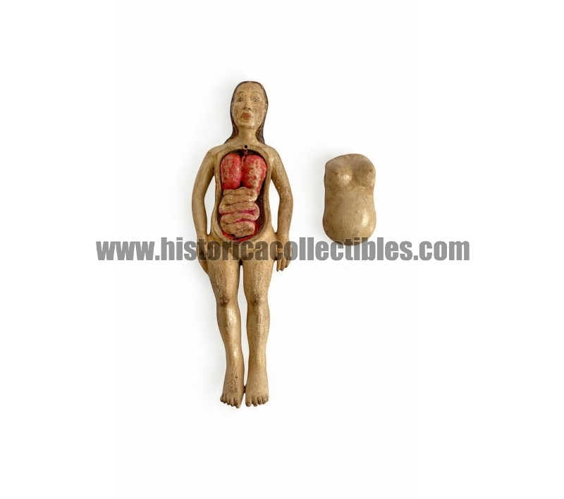
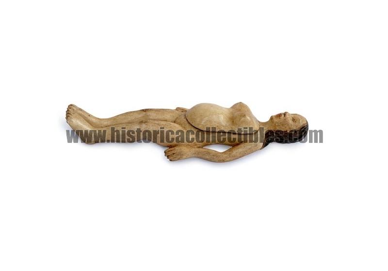

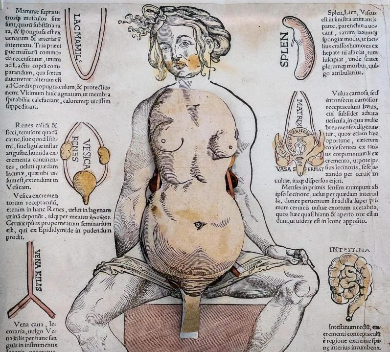
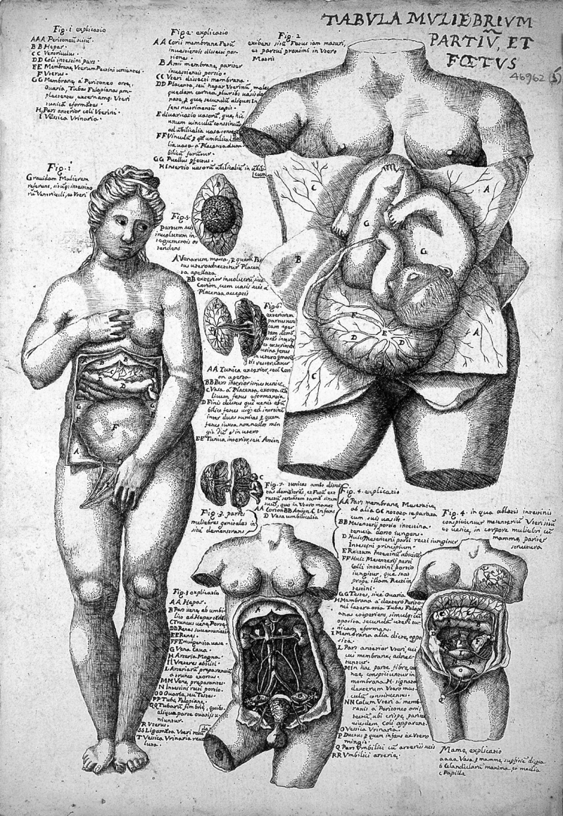
Demonstrative anatomical model of a pregnant woman, in polychrome boxwood, used in lessons to medical students in the second half of the 17th century.
Reclining figure with outstretched arms, loose hair and a high forehead, with the front section of the torso removable to reveal carved models of miniature and removable organs such as lungs, intestines and fetus. Below these are visible, carefully carved and part of the single block of the figure, heart, liver, kidneys and mother's womb, within which there is the space designed to accommodate the fetus.
To underline the preciousness of the handwork, the minute, accurate and only apparently superfluous details that distinguish it should be considered, such as: profile of the nails, pink polychromy of the lips and the different shades of brown used to color the hair.
In light of this, the author of the handwork surprisingly took into account the different shades of red used to color the organs, differentiating the intensity between the more vascularized ones, such as the liver and kidneys, and the less vascularized ones, such as the intestine, for which he used a lighter color.
The level of anatomical detail is limited but provides a basic layout of the main organs. It is therefore likely that these models were made to teach lay people basic human anatomy or they may have been used by midwives to reassure pregnant women and to teach young married couples about anatomy and pregnancy.
Its state of preservation is excellent and it is extremely rare as it is made of boxwood and not ivory.
References: